


02 October 2026
From software updates to multimodal solutions, analytics, and comfort. OT gathers the latest updates in optical coherence tomography (OCT) and imaging.
Take the stage, maestro
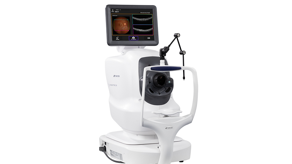
Topcon has launched the Maestro3, a multimodal imaging solution offering 130,000 A-scans/sec, OCT, OCT angiography (OCT-A) and true colour non-mydriatic fundus imaging with a 50-degree field of view – approximately 74.9 degrees at the spherical centre of the eye as confirmed with a model eye.
The device also enables fundus autofluorescence and optional anterior segment OCT imaging.
Alignment, focusing, image capture, and movement from right to left eyes are fully automated with one touch. Some features are optional or only available with the Maestro3 Plus.
Topcon shared that, powered by its IMAGEnet7 Advanced Multimodal Imaging and Analytics, “Maestro3 transforms data into clinical intelligence.”
Combined, this offers a fast, integrated solution delivering “advanced visualisation, quantitative analysis and intuitive reporting to support confident clinical decision making,” the company said.
Enhanced workflow
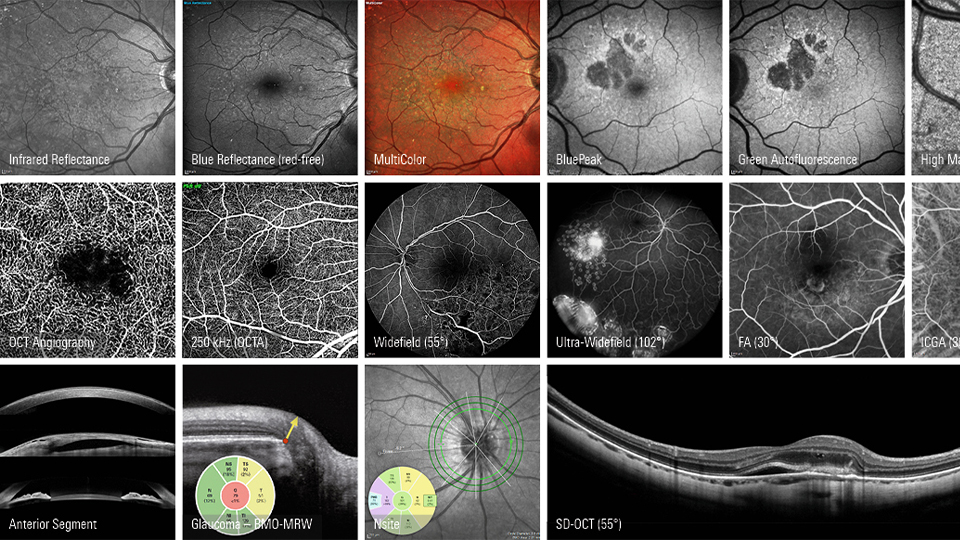
Heidelberg Engineering has released the Spectralis Software 7.4. The software is exclusively compatible with Heyex 2 and is set to enhance workflow efficiency, imaging speed, and diagnostic confidence.
Features include faster 250 kHz scan speed for OCTA imaging with Shift technology. Higher scan speed of 250 kHz with enhanced TruTrack Active Eye Tracking for OCTA reduces acquisition time, the company said, highlighting that shorter examination times can benefit patients who struggle to maintain fixation.
DART (Dense Automatic Real Time) OCTA imaging is a feature, applying ART averaging of up to 100 B-scans to deliver high detail and noise reduction for images of retinal vasculature.
The new Posterior Pole Change Analysis offers flexibility to combine or segment layers in the ganglion cell complex and enables visualisation of patterns of structural change in the macular over time in one view.
Imaging portfolio update
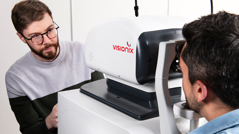
Grafton Optical has been appointed as the exclusive UK and Ireland distributor for Visionix’s advanced Optovue imaging portfolio.
The Optovue Solix is available in two configurations: Solix Essential and Solix Expert, both using ultra-fast spectral domain OCT technology, with FullRange anterior imaging to capture the entire anterior chamber in a single scan.
Solix Essential combines high-resolution anterior and posterior segment OCT with integrates fundus imaging, while Solix Expert adds OCTA.
The newest addition to Grafton Optical’s range is Optovue Autofusion which combines high-resolution, non-contact OCT with an integrated high-resolution fundus camera.
The Optovue iVue80 OCT completes the portfolio – a compact spectral domain OCT designed to provide high-resolution retinal, optic nerve and anterior segment imaging in a space-saving footprint.
Expand the view
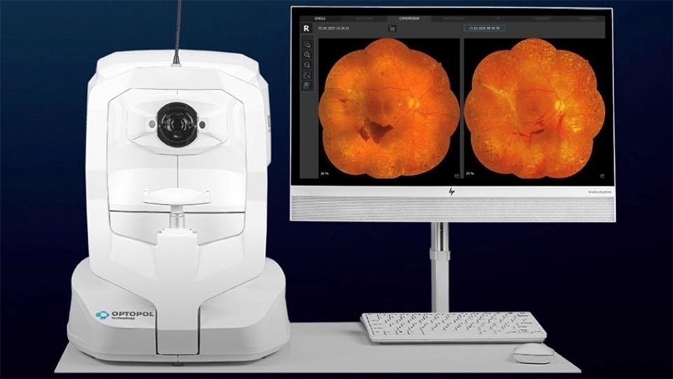
BiB Ophthalmic Instruments has highlighted that the latest software update available on all Optopol Revo FC models facilitates wide-field panoramic true colour fundus imaging.
In Mosaic photo mode, up to nine internal fixation positions allow the capture of peripheral areas and create a wide-field mosaic image without mandatory patient dilation.
The Revo FC now enables coverage of up to 100-degrees (150-degrees at the spherical centre of the eye).
Anterior OCT capabilities
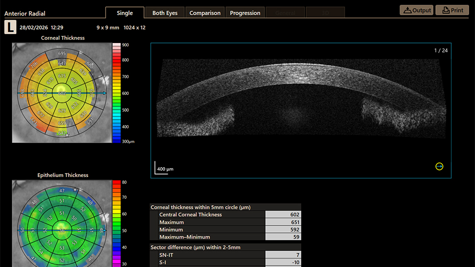
The latest enhancement to the Canon OCT-R1 introduced anterior segment OCT capabilities, supporting earlier and more objective assessment of anterior segment conditions.
Integrating the feature into a single platform enables a more efficient workflow and patient pathway.
Abdul Jahangir, director of Sense Medical, said: “The addition of anterior OCT to the OCT-R1 is a natural progression – bringing greater depth to everyday practice and ensuring clinicians have access to the kind of detailed insight that supports better patient care.”
A broader look
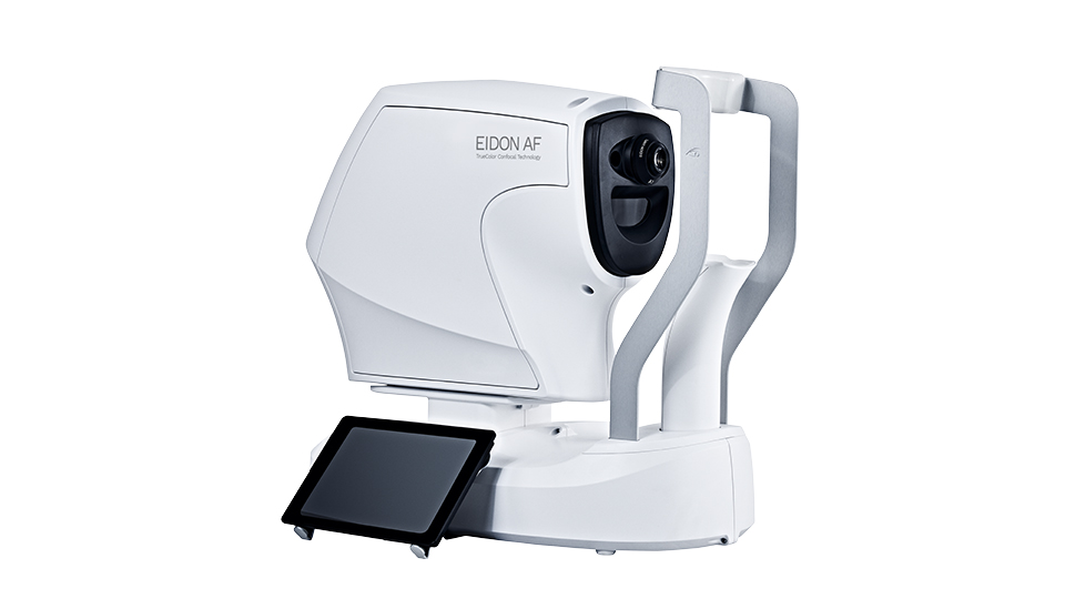
Mainline Instruments highlighted the iCare Eidon which uses TrueColour confocal imaging and captures a single 120-degree wide-field view for a broader look at the retina.
The device is non-mydriatic and uses a gentler light source than traditional flash systems, meaning most patients do not require dilation drops, the company said.
The device can also be configured for autofluorescence and fluorescein angiography. Learn more about the iCare Eidon through OT’s video here.


Comments (0)
You must be logged in to join the discussion. Log in