


13 December 2022
The double-blind, placebo-controlled, monocentric Floater Invention Study (FLIES) examined subjects of all ages over a period of six months and was able to demonstrate significant improvements in subjective visual disturbances as well as in the objective parameters ‘vitreous opacity density’ and ‘contrast sensitivity.’
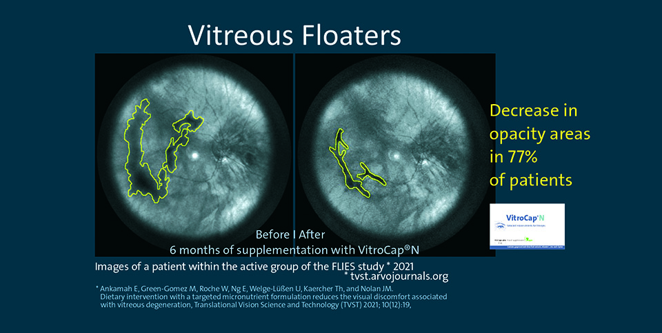
The success rate among the subjects was 77% for the objective improvements (reduction of floater opacity areas) and 67% for the subjective ones (complaint score).
The product used in the study in the verum group was VitroCap®N.
Vitreous opacity and the need of the vitreous body for micronutrients
Vitreous opacities, caused by the aggregation of collagen fibres in the vitreous, appear frequently. They are perceived as spots, specks or thread-like structures and arise from shadows, cast on the retina by the clumped collagen fibres interfering with the incident light. They also cause a reduction of visual functions, detectable in the reduced contrast sensitivity. Vitreous opacities are painless and usually harmless but often lead to serious problems in the form of visual disorders and psychological burdens for those affected.
After analysing the existing literature on the metabolism and micronutrient supply of the vitreous body, the FLIES researchers at the Nutrition Research Centre Ireland (NRCI), which is renowned for its research on the correlation between nutrition and eye diseases, came to the conclusion that oxidative stress is associated with an undersupply of antioxidants from food (similar to other chronic eye diseases such as AMD and cataracts) and subsequent glycation of collagen fibres (crosslinking via sugar molecules). These two processes may be key mechanisms of vitreous degeneration. Conversely, if the supply of micronutrients found in the vitreous body is optimised, an improvement should potentially be detectable.
To test their hypothesis, they chose the scientific gold standard – a prospective, double-blind, randomised supplementation study – and the VitroCap®N capsule as the micronutrient complex to be tested.
The study was published online in October 2021 after a peer review in the ARVO journal TVST (Translational Vision Science and Technology).
Gold standard FLIES study proves subjective and – for the first time – also objective effectiveness of a dietary intervention in vitreous opacity
The FLIES study shows a significant decrease after six months of dietary supplementation with a formulation consisting of 125 mg L-lysine, 40 mg vitamin C, 26.3 mg grape seed extract, 5 mg zinc and 60 mg citrus flavonoids (available as VitroCap®N) in subjective visual symptoms (67%), a significant decrease in vitreous opacity (success rate 76.9%) and a significant improvement in contrast sensitivity compared to placebo.
The study included 61 patients with symptomatic, long-standing vitreous opacities. Randomised and blinded, the subjects took one capsule of the micronutrient supplement (verum) or placebo daily for six months. Before and after the supplementation, various visual function tests (visual acuity, contrast vision) as well as imaging and video examinations of the vitreous to quantify the opacities (optical coherence tomography - OCT, Spectralis®, Heidelberg Engineering GmbH) were carried out.
These objective and subjective clinical findings confirm the original hypothesis that the optimisation of vitreous supply by supplementation with relevant micronutrients with antioxidant and antiglycating properties are suitable to regulate the mechanisms of vitreous degeneration and thereby reduce the visual complaints associated with vitreous floaters.
References
- Ankamah E, Green-Gomez M, Roche W, Ng E, Welge-Lüßen U, Kaercher Th, Barbur J, Nolan JM. Impact of symptomatic vitreous degeneration on photopic and mesopic contrast thresholds, Clinical and Experimental Optometry. 2021, DOI: 10.1080/08164622.2021.1981116
- Mamou J, Wa CA, Yee KMP, et al. Ultrasound-based quantification of vitreous floaters correlates with contrast sensitivity and quality of life. Invest Ophthalmol Vis Sci. 2015;56:1611–1617
- Sebag J, Yee KMP, Wa CA, et al. Vitrectomy for floaters: prospective efficacy analyses and retrospective safety profile. Retina. 2014;34:1062–1068
- Garcia GA, Khoshnevis M, Yee KMP, et al. Degradation of contrast sensitivity function following posterior vitreous detachment. Am J Ophthalmol. 2016;172:7–12
- Garcia GA, Khoshnevis M, Yee KMP, et al. The effects of aging vitreous on contrast sensitivity function. Graefes Arch Clin Exp Ophthalmol. 2018;256:919–925
- Sebag J, Yee KMP, Nguyen JH, et al. Long-term safety and efficacy of limited vitrectomy for vision degrading vitreopathy resulting from vitreous floaters. Ophthalmol Retina. 2018;2:881–887
