


The use of technology in practice improves the efficiency of eye care services and provides significant benefits for patients and the multi-disciplinary team involved in their care. Technology aids with early detection prompting early intervention, documentation of disease progression, accurate referrals, and patient education, as they can visualise and process the information they’re given about their ocular health.
Another thing I found useful was when my supervisor, Caoimhe, would show me scans of patients she had that day and would ask for differential diagnosis and how I would manage that patient. Sometimes, I would not have an answer so I would go away and read up on possible causes and management to discuss with her the next day.
The skill of accurately interpreting scans requires practice and experience, which was challenging as these are both things that come with time. Due to the detailed nature of the information provided, I found it best to start off familiarising myself with simpler scans showing healthy eyes, so when I was presented with an abnormal result, I would be able to spot the anomalies more easily.
I also found it beneficial to be able to interpret images for more common ocular complications, for example, dry vs wet age-related macular degeneration, cystoid macular oedema and vitreomacular traction, before expanding my knowledge to more complex cases. As good as technology is, it has its limitations. It was useful to familiarise myself with the limitations of practice equipment, to allow for realistic expectations.
The skill of accurately interpreting scans requires practice and experience, which was challenging as these are both things that come with time
Aiding pathology
I saw a patient a few months ago who had attended for a routine eye examination with no presenting symptoms, so when it came to performing Volk, I was not expecting to see anything abnormal.However, the patient had two retinal holes in the extreme temporal periphery, one superior and one inferior, with vitreous traction between the two. This was the case in both of her eyes, and was completely unexpected for both the patient and myself.
When I was explaining my findings and management to the patient, I was able to show her exactly what I was talking about. The Optomap proved extremely useful, as it fully documented my concerns and helped me show them to the patient. It also helped me to perform a swift referral.
I referred the patient urgently due to the size of the retinal holes and the accompanying vitreous traction. She returned a few weeks later to update me on the laser the hospital eye service had performed, and was extremely grateful for the services provided.
My practice does not use artificial intelligence currently, however it has been discussed. I think it is something that will be a great tool to improve diagnostics and potentially initiate earlier intervention, as algorithms can be used to aid with early detection of common ocular pathology.
At the time of writing this, I was preparing for my Stage Two assessments, which were both only a few days away. Since then, I have passed both my overarching and direct observation assessments first time. I am now preparing for July OSCES.
Rochelle says...
I am currently working towards... completing my Stage One journey. Once that’s completed, I will be concentrating on studying for Stage Two and focusing more on what clinically interests and challenges me for further learning. I would love to widen my knowledge as much as I can whilst on placement.
I was able to adapt to the clinical technology by... being open minded and versatile with learning a completely new system in a new environment. I hadn’t worked at Vision Express beforehand, so it was a great learning curve, but thankfully welcome sessions were available. My level of learning has become more enhanced with use of the OCT machine, providing myself a more in-depth understanding.
I am most looking forward to... covering my competencies, and having more clinical discussions and interactive sessions at the training days in Nottingham. Hopefully moving into Stage Two shortly, and continuing to deepen my understanding of the various coding languages I’m learning, such as Python and JavaScript, to help me potentially move more into the IT optometry sector in the future.
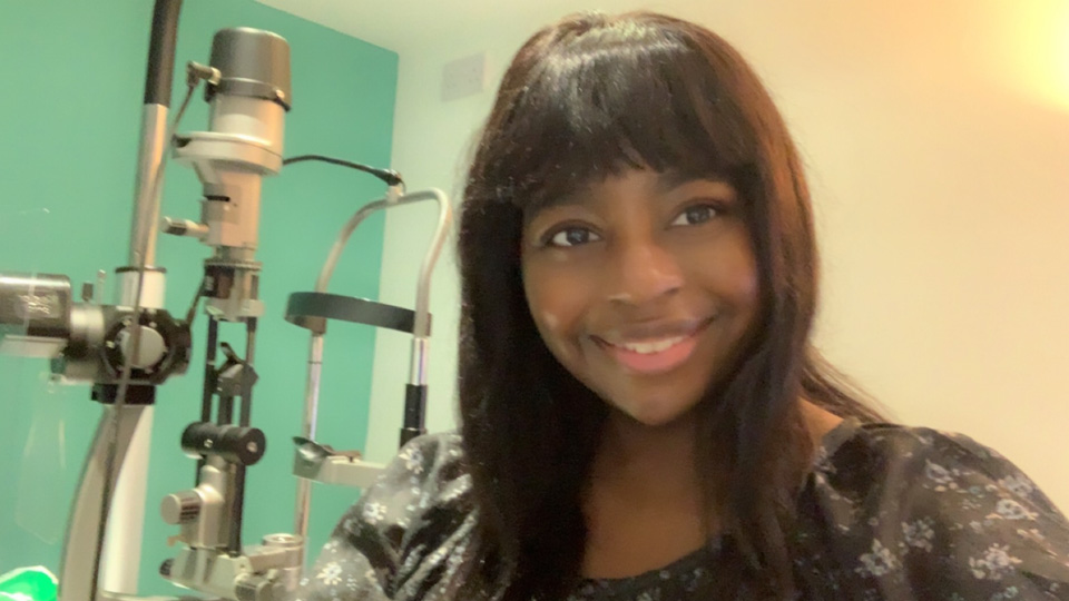
Rochelle Anderson is a pre-registration optometrist at Vision Express Chiswick
I was able to adapt to the clinical technology by... being open minded and versatile with learning a completely new system in a new environment. I hadn’t worked at Vision Express beforehand, so it was a great learning curve, but thankfully welcome sessions were available. My level of learning has become more enhanced with use of the OCT machine, providing myself a more in-depth understanding.
I am most looking forward to... covering my competencies, and having more clinical discussions and interactive sessions at the training days in Nottingham. Hopefully moving into Stage Two shortly, and continuing to deepen my understanding of the various coding languages I’m learning, such as Python and JavaScript, to help me potentially move more into the IT optometry sector in the future.
Rochelle Anderson is a pre-registration optometrist at Vision Express Chiswick
Comments (0)
You must be logged in to join the discussion. Log in