21 December 2017
Two decades since its introduction, optical coherence tomography (OCT) has become indispensable for research, screening, diagnosing, and monitoring diseases of the macula and optic nerve head.
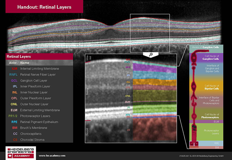
In a high quality, high resolution OCT scan, at least 13 retinal layers can be identified. The inner retina consists of the internal limiting membrane through to the external limiting membrane, and the outer retina consists of the photoreceptor layers through to the choroid. The various layers appear as either bright (hyperreflective) or dark (hyporeflective) bands, depending on whether the layer is reflecting or absorbing light.
"In a high quality, high resolution OCT scan, at least 13 retinal layers can be identified"
It is important to be able to identify and distinguish the different layers of the retina when interpreting OCT scans as identifying an abnormality in a specific layer of the retina can assist the clinician in refining their differential diagnosis. For example, exudates and drusen often look similar with an ophthalmoscope or in fundus photos, but can be differentiated easily based on their location in the retinal layers. Exudates are usually located in or adjacent to the outer plexiform layer because they are lipid residues that originate from damaged capillaries in the inner retina, whereas drusen are deposits between the retinal pigment epithelium (RPE) and Bruch’s membrane because the RPE is not functioning correctly.
In conclusion, it is important to have an appreciation of retinal anatomy and physiology combined with a multimodality imaging approach for the correct management and referral of patients with eye disease.
Visit the Heidelberg Engineering Academy website for a free Retinal Layers Atlas.