- OT
- Industry
- Equipment and suppliers
- Vision's future
Advertorial
Vision's future
What if you could more accurately evaluate a patient's future vision? Right now. In your office
Advertorial content is paid for and produced by a sponsor, and is reviewed and edited by the OT team before publication.
30 May 2017
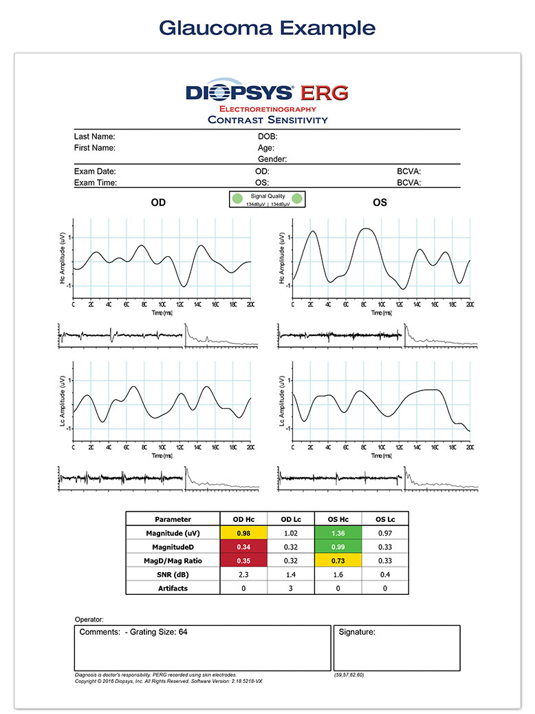
As the world leader in modern visual electrophysiology, Diopsys, Inc. is excited to introduce the clinical benefits of performing electroretinography (ERG) and visual evoked potential (VEP) vision tests right in the eye care practice. Results from these tests are not just for rare disorders, but also for the early diagnosis and management of more common diseases like glaucoma, age-related macular degeneration and diabetic retinopathy. (1–3)
For instance, a study out of Bascom Palmer Eye Institute on glaucoma suspects showed that changes in pattern ERG results can be detected approximately eight years sooner than changes in RNFL thickness, as measured by optical coherence tonometry. The authors conclude: "This represents a substantial window for intervention before permanent loss of structure from glaucoma."1
Diopsys has done more than any other company to advance the use of ERG and VEP in the clinical practice. Visual electrophysiology instruments have historically been expensive and cumbersome, and the tests difficult to perform and interpret. In response to these challenges, Diopsys created an accessible, in-office visual electrophysiology suite, including PERG, VEP, and ffERG vision tests.
To overcome the device and result interpretation issues, Diopsys CEO, Joseph Fontanetta, explained: "We have developed two different practice-friendly testing platforms, the Diopsys® NOVA™ cart system and the Diopsys® ARGOS™ tabletop system. And through our extensive clinical research, we can now provide eye care specialists with clear test results that are colour-coded based on documented reference ranges."

This is modern visual electrophysiology for the eye care practice.
- Clear, intuitive reports
- Non-invasive lid sensors
- Compact, table-top device.
For more information, visit the Diopsys website.
References
- Banitt et al. Progressive Loss of Retinal Ganglion Cell Function Precedes Structural Loss by Several Years in Glaucoma Suspects. IOVS, March 2013, Vol. 54, No. 3
- Oner et al. Pattern electroretinographic results after photodynamic therapy alone and photodynamic therapy in combination with intravitreal bevacizumab for choroidal neovascularization in age-related macular degeneration. Doc Ophthalmol. 2009 Aug;119(1):37-42
- Pescosolido et al. Role of Electrophysiology in the Early Diagnosis and Follow-Up of Diabetic Retinopathy. J Diabetes Res 2015 5;2015:319692.


