

30 January 2017
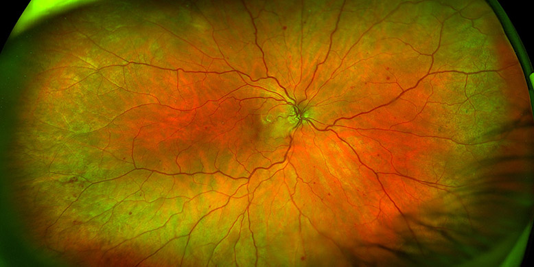
The periphery matters is the assertion that is typically embraced in cases, such as retinal breaks, in which a patient might present with flashes and floaters, and a practitioner is prompted to explore the retina further. However, it is not as commonly understood in the early detection and progression of other pathologies and diseases, which are often asymptomatic in early stages, such as in diabetic retinopathy.
Contact Optos for more information on attending a talk by Jeffry Gerson at 100% Optical
A global perspective
In a 2015 study that compared ultra-widefield (UWF) optomap technology with early treatment diabetic retinopathy study (ETDRS) (7 Standard) film photographs, 40% of diabetic lesions were discovered outside of the ETDRS gold standard area. Furthermore, the presence of the predominantly peripheral lesions were associated with the nearly five times greater progression of diabetic retinopathy over four years, independent of baseline severity and A1C.
An additional study that was undertaken demonstrated that an optomap-assisted fundus examination might enhance the detection of retinal lesions by 30%, when compared with traditional fundus examination alone. In short, without history or symptom, a preponderance of imperative data lurks outside the standard view.
Expanding our view
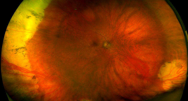
UWF optomap imaging has opened an expansive view of the landscape, exposing otherwise obscured issues. The increasing utilisation of this technology is rapidly changing the vistas of ocular healthcare. Contrasting exploration prompted chiefly on a patient’s report of symptoms or history, and conducted with limited scope, optomap imaging captures a high resolution, 200° digital image of the peripheral retina, as well as the central pole, in a single dynamic image.
However, he stresses that the advances in optomap technology continually enhance how he practises. “Over the years I have witnessed the updates and upgrades as well as the advances in image quality,” he shares, adding: “I believe initially one of the big concerns for practitioners was image quality. That issue really doesn’t exist anymore. The image resolution is excellent.”
"If you are not looking at the peripheral retina, you don't have the whole picture and you are not able to adequately assess risk. This is why we need to look at everybody and see what is out there"
Illuminating patient education
Dr Gerson describes grasping the potential and importance of utilising all of the functions of optomap technology as “an evolving mindset,” wherein practitioners must progressively venture beyond the ability to obtain a UWF colour image and further utilise red/green separations and AF to obtain a comprehensive picture of a patient’s ocular health.
About the author
Jeffry D Gerson is a practising optometrist, as well as a writer, lecturer and thought leader on retinal disease management. He graduated from Indiana University School of Optometry in 1997 and completed his residency at the VA Medical Canter in Kansas City. He went on to become faculty at the University of Kansas School of Medicine. Dr Gerson is a member of American Optometric Association and Kansas Optometric Association, which named him Young OD of the Year in 2008. He is also a fellow of Academy of Optometry and Optometric Retina Society and serves on numerous advisory boards.
Book a place
Watch Jeffry Gerson speak at 100% Optical 2017. His main stage lecture on Why the retinal periphery matters will be held on Sunday, 5 February from 11–12.00.
A workshop on How to successfully implement UWF imaging will be held in the Equipment Hub on Saturday, 4 February, from 13.45–14.45 and Monday, 6 February from 14.45–15.45.
For more information on the talks, contact Optos.