

18 February 2019
3D: diabetic screening
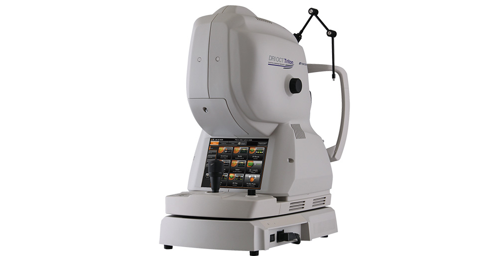
Topcon highlighted its 3D Spectral Domain OCT-1 Maestro and its DRI Triton OCT systems.
The Maestro has an integrated digital colour fundus camera for diabetic screening. It is an automated device that is operated by touchscreen and it has a small footprint.
Topcon’s DRI OCT Triton and Triton Plus models use swept source OCT technology that provides 100,000 A-scans per second, eye tracking and invisible scan lines to minimise patient distraction.
Director of medical sales at Topcon, Graham Trevor, said: “OCT in practice is invaluable. It helps in early detection of changes in various ocular conditions, such as diabetic retinopathy, age-related macular degeneration and glaucoma. The ability to see below the retinal surface with such clarity, ensures confident and accurate referrals.”
The equipment company noted that practitioners investing in OCT should consider post-sale support and training in order to keep informed and maintain confidence when using the device.
For further information visit the Topcon website.
Software: corneal topography
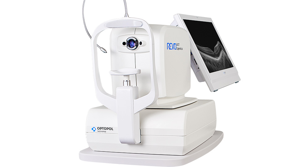
BiB Ophthalmic Instruments has launched the version nine software update for its Optopol Revo Spectral Domain OCT.
The update provides enhanced imagery to assist with more accurate data collection and clinical evaluation.
A new layer has been added to the device to provide detailed corneal topography of the anterior and posterior surface.
The device offers practitioners a high level of accuracy that is ensured by regular calibration. BiB said that this helps practitioners identify corneal changes that may occur due to disease or contact lens interaction.
CEO of BiB, Tim Baker, said: “Optopol are the first to provide T-OCT – the world’s first implementation of a detailed corneal analysis performed by a posterior dedicated OCT.”
BiB explained that with the existing biometry data and anterior analysis, the device offers end-to-end benefits for general contact lens clinics and those who specialise in kerataconic patients and myopia management.
For further information visit the BiB website.
Retina: movie image
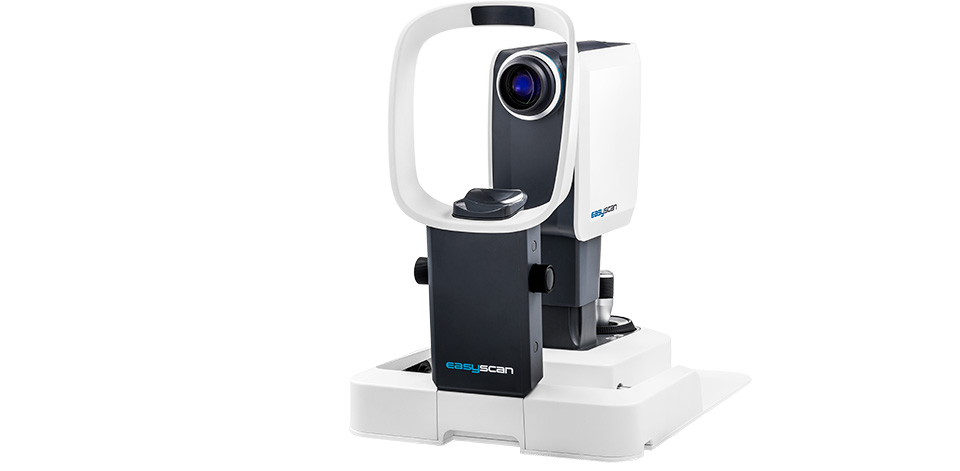
EasyScan highlighted its retinal scanner that utilises scanning laser ophthalmoscope technology to provide scans from the front of the retina to retinal pigment epithelium layers.
The device scans through pupils as small as 1.5mm with no dilation, it can image in a brightly light room and through early stage cataracts.
EasyScan also takes a movie image every time it scans so that practitioners can distinguish between a possible haemorrhage and floaters.
The company explained that its green laser image function is designed for viewing aneurysms, dry age-related macular degeneration and glaucoma. Its infrared laser image is designed for viewing soft drusen, wet age-related macular degeneration and retinal detachment.
UK business development manager at EasyScan, Graham Avery, said: “Both scanning laser ophthalmoscope and OCT purchases are significant investments for any practice, so it’s important to consider what you want to use it for, how it will fit into your workflow process and how quickly it’s going to start earning income for the practice.”
For further information visit the EasyScan website.
Cloud: lens metrology
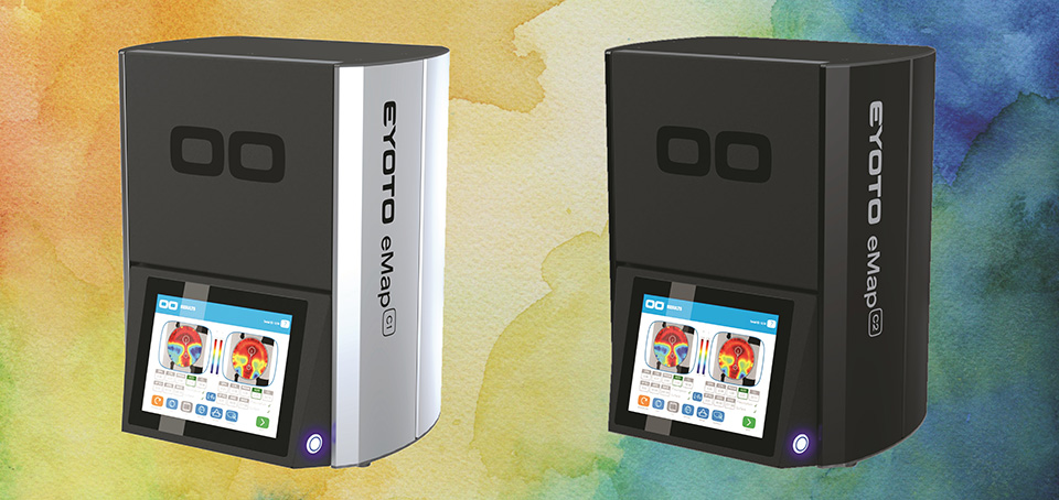
Eyoto launched Cloud Portal at 100% Optical (12–14 January), the next generation cloud system for its eMap C1 and C2 devices.
The company explained that its cloud connect lens metrology system provides objective and repeatable quality control.
It added that the technology provides measurement and quality data to lab managers in real time anywhere in the world.
Chief technology officer and creator of the technology at Eyoto, Dr Thomas Drew, said: “The eMap C1 and C2 represent a step change in technical capability and sets the standard for the small and medium sized lab quality control department. For the first time, lab managers will have total control of the quality control process with unparalleled visibility of data, metrics and productivity all available at their fingertips.”
Eyoto’s eMap C1 images a full, semi-finished or uncut lens by producing a detailed surface power map with prescription confirmation and a visual check for scratches, chips and coating faults.
The C2 device is designed for confirming the prescription of complete edged spectacles, as well as heights and prism tolerance.
For further information visit the Eyoto website.
Diagnostic: ocular surface
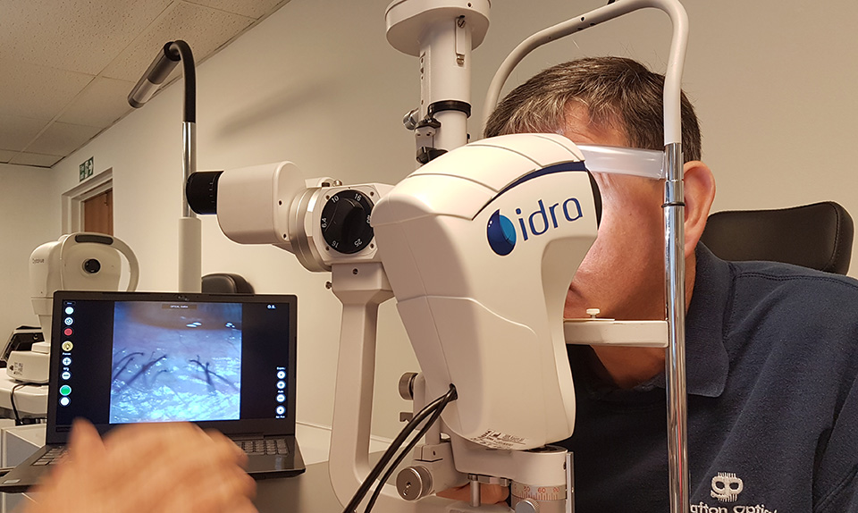
Grafton Optical launched the IDRA Ocular Surface Analyser at 100% Optical (12–14 January).
The company explained that the device is the world’s first comprehensive diagnostic system for high quality tear film analysis.
IDRA provides detailed structural analysis of the lipid, aqueous and mucin layers of the tear film composition.
It enables the user to identify the quality and quantity of a patient’s tear film, as well as the type of dry eye disease present.
Other examinations featured on the device include auto-interferometry, tear meniscus height measurement and auto-non-invasive break up time.
CEO of Grafton Optical, David Thickens, said: “IDRA’s extensive features allow clinicians to show their patients how different treatment approaches can benefit them and demonstrate quantifiable improvements throughout their treatment journey.”
For further information visit Grafton Optical website.
Software: magnified view
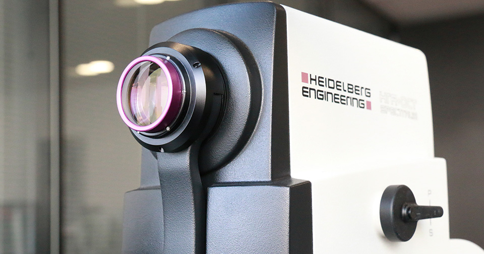
Heidelberg Engineering will launch a high magnification module for its Spectralis OCT device in 2019.
The company explained that the module enables practitioners to see a more magnified and enhanced resolution view of previously unperceivable retinal microstructures.
The ability to see at a microscopic level enables the eye care professional to customise surgical and treatment regimens for the best possible visual outcome, Heidelberg Engineering said.
Head of product management and clinical affairs at Heidelberg Engineering, Ali Tafreshi, said: “The ability to assess ocular microstructures in a magnified view and at an enhanced resolution may provide unique insight into the manner in which diseases progress.”
The lens and software upgrade will be available to new and existing Spectralis diagnostic imaging devices.
For further information visit the Heidelberg Engineering website.
Comments (0)
You must be logged in to join the discussion. Log in