


06 July 2016
Fundus biomicroscopy lenses are probably the most important tool that the majority of optometrists have at their disposal to screen and diagnose posterior eye pathology. Volk is a company that produces these lenses, and such is the popularity of the manufacturer that fundus biomicroscopy lenses are now commonly referred to as ‘Volk’ lenses.
I use these lenses in every clinic when possible, in both private practice and in the hospital setting. As practitioners, we can be guilty of moving too quickly to computerised methods of assessment – optical coherence tomography and fundus photography, for example – but I believe a complete stereoscopic screening evaluation of the vitreous, retina, macula and optic nerve is hard to beat.
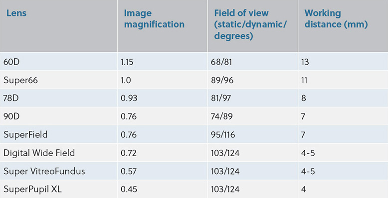
At this moment in time, I routinely use three different Volk lenses – a 90D, a ‘SuperField’ and a 60D lens.
I use a 90D lens mainly on patients with small pupils because it has an 89° dynamic field of view (FOV), 0.76x image magnification and 7mm working distance. While there is a ‘SuperPupil XL’ lens available, I find the 4mm working distance and low magnification difficult to routinely work with.
"As practitioners, we can be guilty of moving too quickly to computerised methods of assessment, but I believe a complete stereoscopic screening evaluation of the vitreous, retina, macula and optic nerve is hard to beat"
The ‘SuperField’ lens is one I use for the vast majority of my patients. It has the same magnification as the 90D, but its 116° dynamic FOV enables it to be an excellent diagnostic lens. The ‘Digital Wide Field’ has a larger FOV, but again, I find that the working distance doesn’t allow a lot of lens manipulation (4–5mm).
For enhanced optic disc and macular assessment, I use a 60D lens. The 1.15x magnification and 13mm working distance makes even the smallest of defects visible. The ‘Super66’ is also a great alternative lens, and has the added benefit of having 1.0x image magnification, meaning no conversion calculations are required to work out the true size of the optic disc or a lesion.
The table below helps to compare some of the lenses features.
As a routine user of this trio of Volk lenses, I have picked up a few tips that help me every day during my clinical examinations that I’d like to share:
Tip one
The red-free filter (green light) can be used to visually enhance lost nerve fibre bundles in glaucoma, or to give greater contrast to haemorrhages and the retinal vasculature.
Tip two
Tilting the lens gives a better view in the far periphery – tilt the lens towards the centre for each direction. For example, with the patient looking down, tilt the superior edge of the lens closer to the eye.
Tip three
Carry out an internet search on ‘Watzke-Allen test’ to help with macular hole assessment.
Tip four
If the patient is significantly light-sensitive, moving the illumination system very slightly off axis helps the patient tolerate the light better, while also enabling you to retain a stereoscopic image. If a macula assessment is proving difficult due to light sensitivity, reduce the slit beam to 0.2mm (a round spot). Although this can make the assessment difficult, the patient should be okay with it, even with the light on the brightest setting.
Tip five
I would never substitute high quality Volk lenses with cheaper alternatives. They do not have the patented double aspheric glass that Volk uses, resulting in much poorer image quality.
Comments (0)
You must be logged in to join the discussion. Log in